
How to report a CBCT scan: a step-by-step guide with a free report template
Every CBCT scan must have a written clinical evaluation covering the whole scanned volume, not only the tooth or site you were interested in. This guide sets out who can report a CBCT scan in the UK, a systematic way to review the volume, what to look for in each region, and how to write the findings up. A free CBCT report template is included further down.
It is written for dentists, specialists and dental care professionals who take or refer for cone beam CT. If you would prefer a radiologist to report your scans, 3Beam offers a CBCT reporting service for scans taken at your practice.
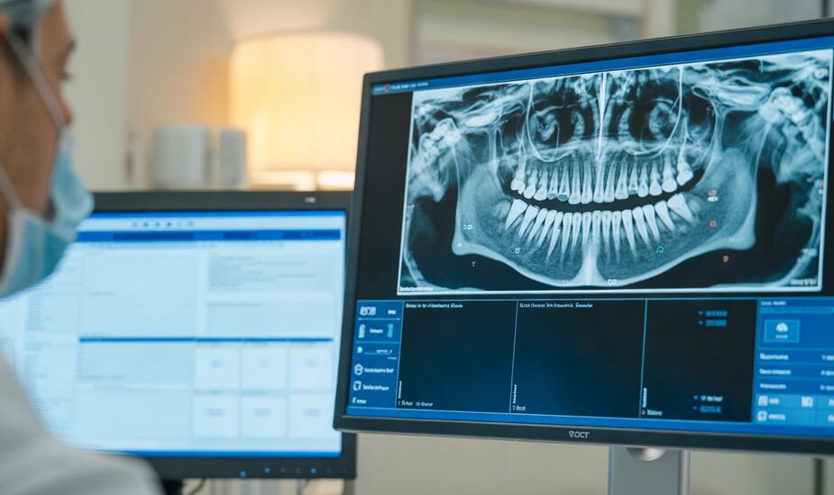
Who can report a CBCT scan in the UK?
A CBCT scan can be reported by anyone who is adequately trained and entitled by their employer to do so, but the scope depends on what the scan covers.
The legal duty. Regulation 12(9) of the Ionising Radiation (Medical Exposure) Regulations 2017 requires the employer to make sure a clinical evaluation of every exposure is carried out and recorded. For CBCT, that evaluation is the written report.
The whole volume. The European SEDENTEXCT basic principles state that the clinical evaluation must cover the entire image dataset. A report that covers only one tooth or one implant site is incomplete.
The referrer's position. If you refer a patient for a scan and the images come back without a report, you become the reporting operator. The HPA and BSDMFR core curriculum is explicit that you must be adequately trained for that task.
| What the scan covers | Who should report it |
|---|---|
| Small volume, dento-alveolar: teeth, supporting structures, mandible, and maxilla up to the floor of the nose (roughly 8 cm x 8 cm or smaller) | A dentist with further (Level 2) training in dento-alveolar CBCT interpretation, or a dental and maxillofacial radiologist |
| Anything beyond the dento-alveolar region: TMJs, sinuses, skull base, temporal bone, cervical spine, and all craniofacial volumes | A dental and maxillofacial radiologist or a clinical radiologist |
UK training expectations are set out in the Guidance Notes for Dental Practitioners on the Safe Use of X-ray Equipment (2nd edition, FGDP(UK) and Public Health England, 2020). Core (Level 1) training covers referrers and operators. Further (Level 2) training is for dentists who justify and interpret dento-alveolar scans.
If a scan includes structures you are not trained to assess, the whole volume still has to be reported. Send it to a radiologist.
Before you start: five checks
A report is only as good as the information and images behind it, so check these before you open the first slice.
The clinical question. Read the referral. You need the reason for the scan, the relevant history and the specific question to be answered.
Previous imaging. Have earlier radiographs or scans to hand so you can comment on change.
The full dataset. Report from the complete DICOM volume in a multiplanar viewer, never from screenshots or a single panoramic reconstruction.
Scan details. Note the date of the scan and the field of view. These go in the report.
Image quality. Look for movement, metal artefact and noise. If they limit what you can see, say so in the report.
Remember what CBCT cannot do. Soft tissue contrast is poor, and grey values are not reliable Hounsfield units, so do not use them to characterise a lesion or to quote bone density.
How to read a CBCT scan systematically
Use the same sequence for every scan, so that nothing in the volume goes unreviewed. Findings outside the area of interest are the easiest to miss.
- Confirm identity and orientation. Check the patient details and the left and right markers. Align the volume so the planes are true to the patient, not to how they sat in the machine.
- Scroll the whole volume in the axial plane. Go from the top slice to the bottom, slowly, looking at everything.
- Repeat in the coronal and sagittal planes. A finding should be confirmed in at least two planes before you call it.
- Build the reconstructions you need. Draw a panoramic curve through the arch and step through the cross-sections for each jaw.
- Examine the region of interest in detail. Use thin slices, adjust brightness and contrast, and take the measurements the referrer asked for.
- Review every other structure in the field. Work through the region checklist below.
- Compare with previous imaging. Record whether findings are new, stable or changing.
- Return to the clinical question. Make sure the report answers it directly.
CBCT reporting checklist: what to look for in each region
Work through every region that appears in the field of view, and record a finding or a normal statement for each one.
| Region | What to assess | Do not miss |
|---|---|---|
| Teeth | Number, position, impactions, root form, canal anatomy, root fillings, resorption, fractures | Untreated canals, perforations, external cervical resorption |
| Periapical tissues | Periapical radiolucencies, widened ligament space, sclerosis | A lesion larger or less defined than the clinical picture suggests |
| Alveolar bone | Bone levels, furcations, bone height and width at edentulous sites, cortical plates | Dehiscence or fenestration at a planned implant or orthodontic site |
| Mandible | Inferior alveolar canal course, mental foramen, anterior loop, lingual vascular canals, lingual concavity, bone lesions | Bifid canals, cortical breach, an ill-defined lesion |
| Maxilla | Incisive canal, sinus floor, tuberosity, residual ridge | Sinus floor breach, oroantral communication |
| Maxillary sinuses and nasal cavity | Mucosal thickening, opacification, fluid levels, retention cysts, septa, ostium, nasal septum, turbinates | Bony wall erosion, complete unilateral opacification |
| Temporomandibular joints | Condylar shape and cortex, erosions, osteophytes, joint space, symmetry | Destructive change, marked asymmetry |
| Airway | Width and symmetry of the nasopharynx and oropharynx, adenoids, tonsils | An asymmetric soft tissue mass, marked narrowing |
| Cervical spine | Alignment, vertebral body shape, degenerative change | A lytic or sclerotic lesion, loss of alignment |
| Skull base and other bone | Styloid processes, mastoid air cells, any skull base in the field | Bone destruction, opacified mastoid air cells |
| Soft tissue calcifications | Carotid artery, salivary glands, tonsils, lymph nodes, stylohyoid ligament | Carotid artery calcification, which needs referral to the patient's GP |
Incidental findings are common on CBCT. Most are harmless, but some need medical or surgical follow-up, so each one should be recorded and acted on.
How to write a CBCT report
A good CBCT report answers the clinical question, describes every relevant finding precisely, and tells the referrer what to do next.
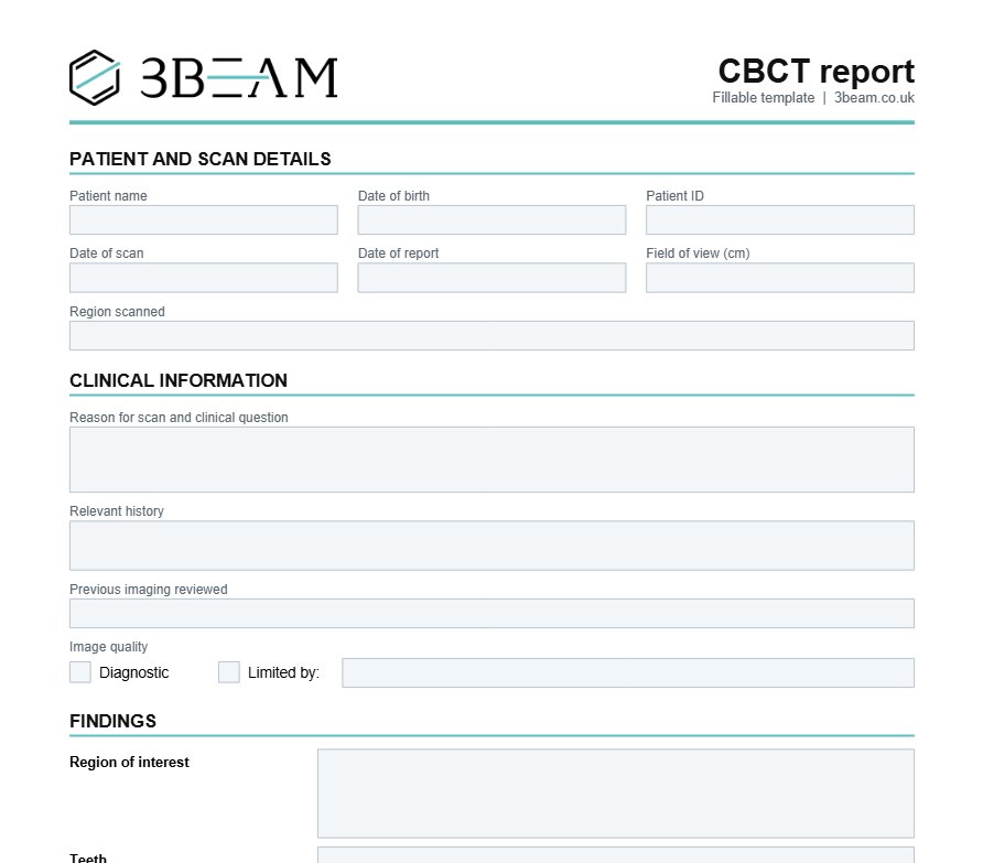
What a CBCT report should include
- Patient details, date of scan and date of report
- Field of view and region scanned
- The clinical information and the question asked
- A statement on image quality and any limitations
- Findings, region by region, including incidental findings
- Measurements, where requested
- A conclusion that answers the question
- Recommendations, including any onward referral
- The reporter's name, qualifications, registration number and signature
How to describe a lesion
Describe it in the same order every time: site, size, shape, margins, internal structure, and effect on adjacent structures.
- Site: name the tooth or region and the side, using one tooth notation throughout.
- Size: give three dimensions in millimetres.
- Margins: well defined and corticated, well defined and non-corticated, or ill defined.
- Internal structure: radiolucent, radiopaque or mixed.
- Effects: root resorption, tooth displacement, cortical expansion, thinning or perforation, canal displacement.
Wording that helps the referrer
- Give a differential diagnosis in order of likelihood, not a list of every possibility.
- State what you could not assess and why, for example metal artefact around a restoration.
- Take ridge measurements perpendicular to the ridge, and state where each was taken.
- Record normal findings for each region reviewed. Silence reads as "not looked at".
- Telephone the referrer about anything urgent, and record that you did.
CBCT report template
Use this template for every scan and complete every row. Delete a region only if it is not in the field of view, and say so.
Common CBCT reporting mistakes
These are the errors to guard against, starting with the one that carries the most risk: reviewing too little of the scan.
- Reporting only the region of interest. The duty covers the whole volume, including the sinuses, airway and spine if they are in the field.
- Reporting from the panoramic reconstruction. It is a summary view. Findings must be confirmed on the axial, coronal and sagittal slices.
- Mistaking artefact for disease. Dark bands beside metal restorations and root fillings can mimic caries, fractures and bone loss.
- Trusting grey values. They vary between machines and across the volume, so they cannot be used as bone density.
- Measuring on an oblique slice. A cross-section that is not perpendicular to the ridge overstates bone height and width.
- Missing a second canal. Bifid mandibular canals and accessory foramina are easy to overlook at implant and third molar sites.
- Recording an incidental finding and doing nothing. Tell the patient, refer if needed, and record both.
When to refer the scan to a radiologist
- The field of view extends beyond the teeth and jaws.
- A lesion is ill defined, destructive, or has changed since the last scan.
- You can describe a finding but cannot name it.
- You have not completed further training in CBCT interpretation.
A finding that could be malignant should be referred on your local urgent suspected cancer pathway without waiting for further imaging.
Prefer a radiologist to report your CBCT scans?
3Beam reports CBCT scans taken at your own practice for a fixed fee of £130 per scan whatever the field of view.
- Who reports: consultant dental and maxillofacial radiologists.
- Turnaround: 5 working days as standard, or 24 hours for urgent requests at an extra £50.
- What you receive: a full-volume report with measurements, annotated screenshots and clear findings for treatment planning.
- How to send a scan: upload the DICOM data through our secure form from a desktop computer.
- Contract reporting: available for practices and NHS services that want every scan reported.
Send a scan for reporting or see sample CBCT reports and pricing.
If you would like to build your own reporting skills, see our online CBCT course. You can also refer a patient for a CBCT scan at 86 Harley Street.
3Beam is a CQC-registered provider. Call 0207 637 8227 or email info@3beam.co.uk.
CBCT reporting FAQs
Does every CBCT scan need a report?
Yes. IR(ME)R 2017 requires a recorded clinical evaluation of every exposure, and for CBCT that means a written report covering the whole volume.
Can a dentist report their own CBCT scans?
Yes, for small dento-alveolar volumes, provided the dentist has completed further training in CBCT interpretation and is entitled to report by their employer. Larger volumes should go to a radiologist.
Do I have to report areas outside my region of interest?
Yes. Everything captured in the field of view must be evaluated, including the sinuses, airway, joints and cervical spine where they appear.
Who is responsible for the report if the scan was taken elsewhere?
If the imaging centre supplies a report, its reporter is responsible for it. If the images are returned to you without one, you take on the reporting role.
What should a CBCT report include?
Patient and scan details, the clinical question, image quality, findings by region, incidental findings, a conclusion, recommendations, and the reporter's name and registration number.
How long does a CBCT report take?
3Beam returns reports within 5 working days, or within 24 hours for urgent requests.
How much does a CBCT report cost?
3Beam charges a fixed fee of £130 for any size of CBCT scan. Urgent 24-hour reporting costs an extra £50.
References
- The Ionising Radiation (Medical Exposure) Regulations 2017, regulation 12 (SI 2017 No. 1322)
- Guidance Notes for Dental Practitioners on the Safe Use of X-ray Equipment, 2nd edition, FGDP(UK) and Public Health England, 2020
- SEDENTEXCT basic principles on the use of dental cone beam CT
- Core Curriculum in Cone Beam Computed Tomography for Dentists and Dental Care Professionals, Health Protection Agency working party and BSDMFR, 2009
- Selection Criteria for Dental Radiography, 3rd edition
- Makdissi J, Loughlin A, Dungarwalla MM. Do something or do nothing? Anatomical variants and incidental findings on cone beam CT. Part 1. Dental Update 2022; 49(2): 95-101
- Harvey SC. What to include in CBCT reports. DDU Journal 2026; issue 32
This guide is for dental and medical professionals and is general information, not a substitute for formal training in CBCT interpretation.