
CBCT Neck Pain Scans in Chiropractic
Upper Cervical CBCT Scan in London
At 3Beam Imaging Centre, Harley Street, we are proud to be the UK’s only specialist Cone Beam CT (CBCT) imaging facility capable of performing upper cervical (neck) spine scans. Our state-of-the-art Planmeca CBCT system delivers ultra-high-resolution 3D images of the C1 and C2 vertebrae (atlas and axis) — ideal for chiropractors, osteopaths, and medical professionals requiring detailed cervical imaging.
Cervical CBCT Scan Price
-
Price: £200
-
Reporting: Images only (no radiology report included)
-
Results: All CBCT scan images are securely sent to the referring chiropractor or clinician for interpretation.
Same-Day Appointments Available
We offer same-day appointments for cervical spine CBCT scans to ensure prompt and convenient access to imaging.
Your scan will be performed by Ali Samani (HCPC: RA61962), our highly experienced radiographer and former Deputy Superintendent at Guy’s Hospital, who has extensive expertise in upper cervical imaging.
Scans can be carried out with the patient standing or sitting, and we can accommodate different neck positions, including:
-
Neutral
-
Oblique
-
Rotational
This allows for precise evaluation of alignment and better clinical correlation with patient posture and neck movement.

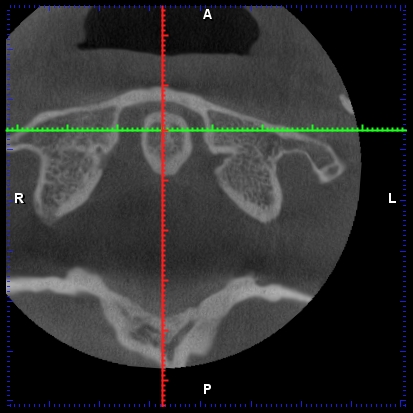
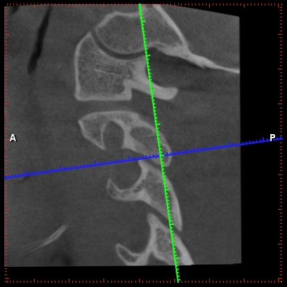
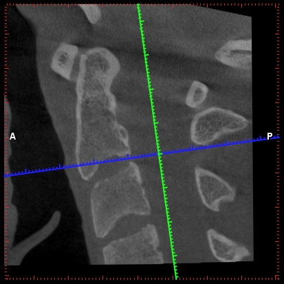
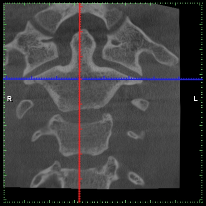
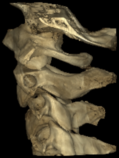
CBCT imaging and upper cervical region
CBCT (cone beam computed tomography) was first developed in dentistry to image the jaw in fine detail for implant planning. That same precision is now transforming how the upper neck is assessed — and it offers a level of detail that standard X-ray simply cannot match.
A CBCT cervical spine scan produces a high-resolution 3D image of the neck and the cranio-cervical junction (CCJ) — the critical area where the skull meets the top of the spine. This makes it a valuable investigation for patients experiencing persistent neck pain, chronic headaches and migraine, and suspected cervical misalignment, where precise visualisation of bony alignment is essential.
Why the cranio-cervical junction matters
The CCJ is one of the most important and vulnerable regions of the spine. Around a third of cervical spine traumas occur at this junction, and injury here can affect the alignment of the upper neck, with potential neurological and vascular implications. Because the CCJ sits so close to the brainstem, it has drawn growing clinical interest in the assessment of patients with post-concussion symptoms and unexplained headache. Detailed 3D imaging allows these structures to be evaluated far more accurately than with conventional radiographs.
The benefits of CBCT for the cervical spine
A CBCT cervical spine scan can help to:
- Visualise the bony alignment of the upper neck and CCJ in three dimensions
- Investigate the structural contributors to neck pain, headaches and migraine
- Assess suspected cervical misalignment with millimetre precision
- Provide detail that supports onward clinical and chiropractic assessment
Crucially, our cervical spine scans are performed in a seated or standing (weight-bearing) position, reflecting the spine's natural upright orientation — unlike conventional CT performed lying down. This gives a more representative picture of how the cervical spine behaves under everyday load.
The radiation exposure is much less than standard X-ray series. A standard CBCT scan is 240 micro sieverts, which is equivalent to about 1/10th the radiation exposure of a standard 4-view cervical spine X-ray series. This is equivalent to the exposure of a 5-hour airplane flight (due to less shielding from cosmic radiation at higher altitudes!)
Since the human structure is 3-dimensional, it is vital to view it with 3D imaging in order to accurately evaluate the alignment. Traditional X-ray is 2-dimensional, so certain factors like anatomical variations, positioning errors and asymmetry of structures can result in misdiagnosis some of the time.
The cranio-cervical junction (CCJ) plays an important role in the overall motion of the cervical spine, accounting for 25% of the flexion and extension, and up to 50% of the axial rotation of the neck.
Upper neck cervical CBCT is used to identify density of bone, fractures, skeletal maturation, cervical fusion assessments, cervical and temporomandibular joint instabilities, atlanto-occipital instability, bony alignment and styloid pathologies.
The detail of bony anatomy provided by a CBCT image of the CCJ is useful to appreciate the components of the CCJ and its role in cervical spine stability and clinical implications.
The CCJ is an area of unique consideration given bone morphology, biomechanics, ligamentous structures, fluid dynamics, neurology, and neurophysiologic effects attributed to that area.
Additionally, CBCT may be indicated to evaluate for traumatic fractures and hairline fractures within the cervical spine. It may also be used to evaluate cervical spine spondylosis, intervertebral foraminal stenosis, and facet joint arthrosis. These abnormal findings are regularly evaluated for in chiropractic offices that specialise in the cranio-cervical junction.
Why Choose 3Beam for Cervical (Neck) CBCT Scans
- Specialist imaging centre dedicated to dental and cervical CBCT
- Only UK facility experienced in upper cervical scanning
- Flexible positioning: standing, sitting, oblique, or rotated
- Same-day availability for urgent cases
- Convenient Harley Street, London location
- Secure digital delivery of scan results to your chiropractor