CBCT in Endodontics
Morita Veraview X800 CBCT scans
Our Morita Veraview X800 CBCT allows us to image the inside, and around your tooth in 3D. This enhances diagnosis as studies have shown 30%-40% of abscesses cannot be visualised through standard 2D x-rays. Fractures and missed canal anatomy can also be identified bringing increased clarity to treatment planning and the chances of successful treatment.
We have equipped our imaging centre with Morita Veraview X800 manufactured in Japan, designed to be the world's first "Total Performance Imaging" system, providing outstanding image quality ensuring a precise and reliable diagnosis in endo practice.
The advantages of Morita CBCT in endo:
· In cases where anatomic superimposition of roots or areas of the maxillofacial skeleton is impeding diagnosis
· Determine the extent of the periapical disease
· In cases where complications have arisen to determine appropriate treatment planning to resolve such issues as:
01 overextended root canal obturation
02 material separated endodontic instruments
03 calcified canal identification
04 localisation of perforations
· Diagnosis and management of dento-alveolar trauma, root fractures, luxation, displacement of teeth, and alveolar fractures.
· Diagnosis of internal/external root resorption or invasive cervical resorption
· Surgical planning needed to determine the location of root apices and to evaluate the proximity of adjacent anatomical structures

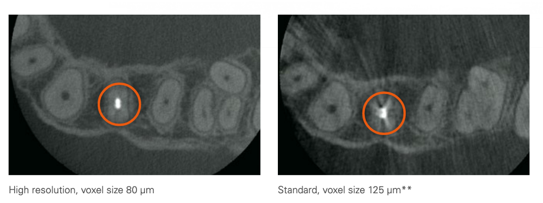
Morita CBCT uses state-of-the-art 3D X-ray technology in its Veraview X800, which provides brilliant image quality with a voxel size of 80 µm and resolution of 2.5 LP/mm and MTF of 10 per cent. Highly precise 3D, panoramic and cephalometric images — both in 180° and 360° modes — can be obtained with this unit.
Furthermore, the wide spectrum provided by 11 fields of view covers many fields of dentistry and is particularly applicable in endodontics.
In order to further increase the sharpness of the images and to reduce artefacts and distortion to a minimum, Veraview X800 uses a horizontal X-ray beam that generates highly detailed 3-D images. If the operator shifts the horizontal X-ray beam by 5°, the disruptive shadows on the hard palate are suppressed in panoramic imaging. Afterwards, the X-rays can be recalculated with the zoom reconstruction function; for example, an 80 μm voxel image can be reconstructed from a 125 μm voxel image without having to take new scans. The new features of Veraview X800 are combined with the R100 Reuleaux triangular field of view, which scans only the region needed for making the diagnosis and, thus, significantly reduces radiation exposure compared with the conventional cylindrical shape.

Dental X-Ray Radiation Risk, Background Radiation and CT machines
X-rays are present in the atmosphere (cosmic radiation) and background radiation also comes from geological structures. Medical x-ray machines produce significant amounts of radiation and the principle governing rule to exposure is to always keep this as low as reasonably possible, employing a favourable risk-benefit ratio.
The level of risk varies according to age and the parts of the body exposed to the radiation. Some areas are more sensitive to others. Dental x-rays generally carry less risk as the jaws are less sensitive areas. The table below (www.xrayrisk.com) calculates the additional risk of cancer from a 4x4cm 3D CBCT scan to an average 40-year-old female. The baseline risk of a cancer is 37.5% (1 in 3) for women and a scan will add a further risk of 0.000285% (1 in 350,887). That is one hundred times less risk than being hit by a meteorite (1 in 3,200).
As a comparison consider that the estimated lifetime risk of being killed in a road traffic accident in the UK is 0.42% (1 in 240 chance) or by a bolt of lightning is 0.001% or 1 in 100,000 chance.
In terms of the amount of radiation, or the dose, it might be useful to compare the doses of various sources of radiation. From dosimetry carried out on our scanner the calculated average dose of the smallest scan was 0.028mSv (Morita X-ray Dosage Information). Based on these figures a 4x4cm Morita scan is equivalent to 4 days of background radiation living in London or just over 1 day living in Cornwall.
| Source of exposure | Dose |
|---|---|
| Dental x-ray | 0.005 mSv |
| 100g of Brazil nuts | 0.01 mSv |
| Chest X-Ray | 0.014 mSv |
| Transatlantic Flight | 0.08 mSv |
| UK annual average radon dose | 1.3 mSv |
| CT scan of the head | 1.4 mSv |
| UK average annual radiation dose | 2.7 mSv |
| USA average annual radiation dose | 6.2 mSv |
| UCT scan of the chest | 6.6 mSv |
| Average annual radon dose to people in Cornwall | 7.8 mSv |
Taken from Ionising radiation: dose comparisons
The radiation exposure doses below are taken from an article in the International Endodontic Journal*
*Patel S, Dawood A, Pitt Ford T, Whaites E. The potential applications of cone beam computed tomography in the management of endodontic problems. International Endodontic Journal, 40, 818–830, 2007
