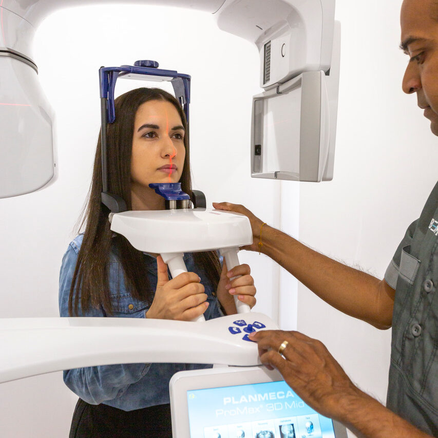
OPG scan (panoramic X-ray)
An OPG is a panoramic X ray of the upper and lower jaws, including the teeth. The unit is specifically designed to rotate around the patients head during the scan, which would take approximately 20 seconds.
An OPG can be used to look for:
- Dental fractures
- Dislocated jaw
- Infection
- Face trauma and fractures
- Impacted wisdom teeth
- Dentition (teeth)
- Dental evaluation for caries or pulp disease
- Trauma assessment for tooth or jaw fractures (broken tooth Xray)
- Sinusitis, periodontitis or periapical abscesses
- Tumour or radicular cyst
- Temporomandibular joint (TMJ) assessment for disease, fractures or dislocations
- Progressive evaluation of orthodontic treatment
An OPG also demonstrates the number, position and growth of all the teeth including those that have not yet surfaced or erupted through the gum. The scan can be particularly useful to check and to see areas like wisdom teeth for impaction, or the development of a child’s jaw and teeth. It is often used to check your TMJ (temporomandibular joint), sometimes called the CMA (cranio-mandibular articulation) especially if you grind your teeth.
The principle advantage of panoramic images / OPGs:
- Broad coverage of facial bones and teeth including the TMJ (temporomandibular joint)
- Very low patient radiation dose
- Convenience of examination for the patient if strong gag reflex present
- Ability to be used in patients who are restricted in opening their mouth
Register here and refer for OPG X-Ray (for healthcare professionals only)
If you are a patient, call us on 0207 637 8227 to discuss your requirements for an Xray
About the procedure
The OPG will be carried out by a radiographer (a health professional trained to perform imaging procedures). They will explain the procedure and make sure that you’re happy to go ahead with it.
- The radiographer will ask you to remove any jewellery in the head and neck area.
- You will be asked to stand in front of the scanning machine.
- A peg will be put into a slot on the machine and you’ll be asked to bite onto this.
- Below the peg are two handles which you’ll need to hold to keep your balance as we may need to ask you to lean backwards to get the best possible picture.
- When you are in the right position, the radiographer will use a clamp to gently hold your head in place and stop it from moving.
- This doesn’t hurt. Once you are in place, the machine starts to move around your head.
- While it is moving it can touch your shoulders. Try not to move as this will blur the image and may mean the procedure has to be repeated. The test usually takes about 15 minutes

