
The new standard for complex endodontic and RC treatment
For modern endodontics treatment and complex root canal treatment, conventional 2D radiographs are increasingly not enough. Curved canals, C-shaped anatomy, persistent periapical pathology and suspected vertical root fractures all demand three-dimensional information if you want predictable outcomes and fewer surprises.
Cone beam CT (CBCT) has become an essential tool in contemporary root canal and endodontic treatment workflows, giving you accurate 3D visualisation of root canal anatomy, surrounding bone and pathology. However, not all CBCT systems are equal – especially when you are dealing with fine endodontic detail.
For endodontics and root canal retreatment, image quality, small field of view and dose are critical. This is where the Morita Veraview X800 stands out.
Why CBCT matters in modern RC treatment
CBCT transforms how you diagnose and plan root canal treatment:
-
Identifies additional canals (e.g. MB2, distolingual canals, C-shaped canals) that are hidden on periapicals.
-
Shows complex root morphology: bifurcations, joins, hooks and curvatures that affect instrumentation and obturation.
-
Improves assessment of periapical lesions that may be underestimated or completely invisible on 2D images.
-
Helps you understand non-healing root canal treatments – missed canals, perforations, cracks or non-endodontic pathology.
-
Supports investigation of vertical root fractures and resorption.
In other words, CBCT provides the extra information that can fundamentally change your endodontic treatment plan – whether you proceed with root canal therapy, attempt retreatment, consider apical surgery or decide to extract.
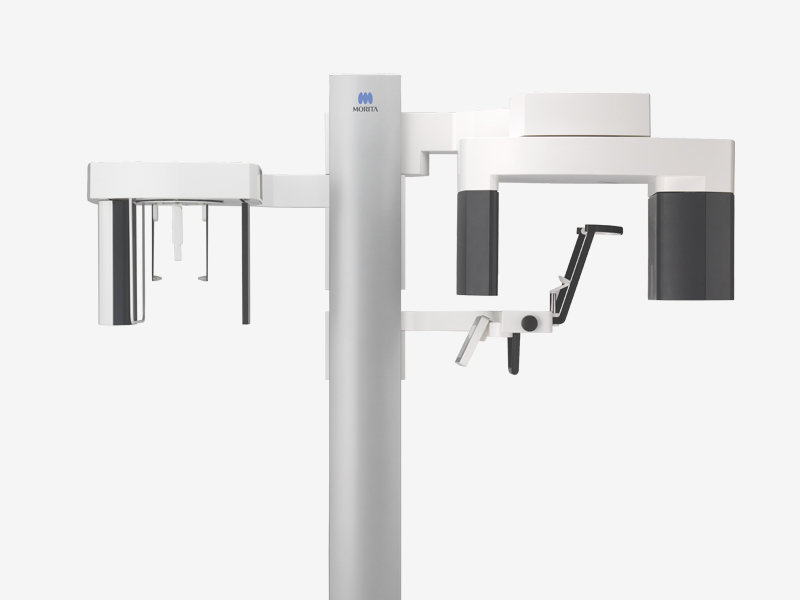
What makes the Morita Veraview X800 different?
For endodontic and root canal indications, you want:
-
The smallest possible field of view
-
The highest useful resolution
-
Low radiation dose and excellent artefact control
The Morita Veraview X800 is specifically designed around these principles:
-
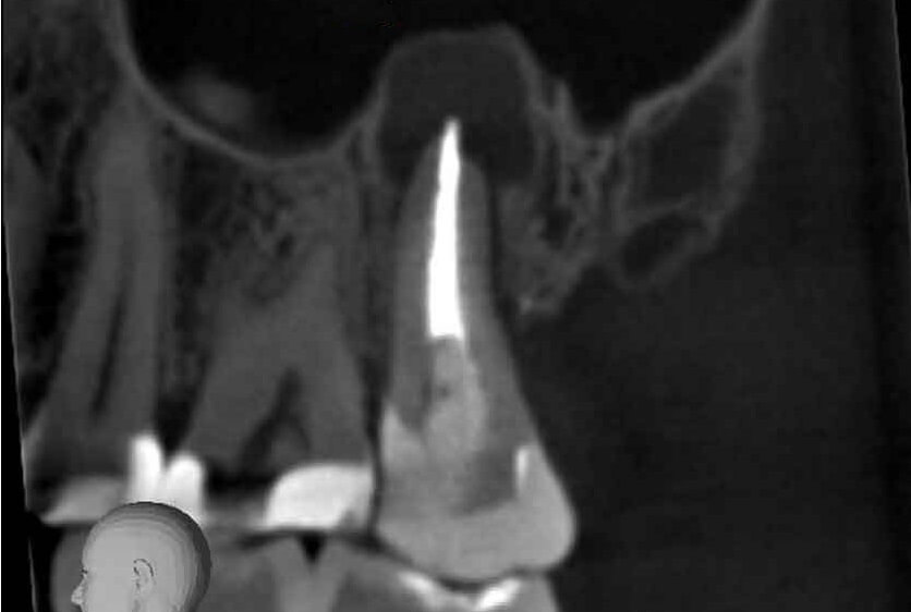
Ultra-high resolution for endodontics
-
Voxel size down to 80 μm in a 4 × 4 mm field of view – ideal for visualising fine canal systems, cracks and small periapical changes.
-
High spatial resolution so you can clearly differentiate small structures relevant to root canal treatment decisions.
-
-
Small, selectable fields of view
-
Multiple small FOV options targeted to single-tooth and quadrant imaging support the “smallest FOV for the clinical question” approach recommended in endodontics.
-
-
Dose-efficient protocols
-
A choice of 360° and 180° scan modes and optimised exposure parameters allow you to balance dose against the level of detail required for a particular root canal case.
-
-
Advanced noise and artefact reduction
-
Morita’s reconstruction algorithms help reduce scatter and metal artefacts from posts, cores and existing root fillings, which is crucial in retreatment cases.
-
This combination of small FOV, high resolution and low artefact is exactly what you need for demanding endodontic treatment planning.
Clinical advantages for your root canal cases
When you refer to a CBCT centre equipped with a Morita X800, there are several clear benefits for your endodontic and root canal workflow.
1. Accurate canal morphology before you start root canal treatment
High-resolution CBCT on a Morita X800 allows you to:
-
Confirm the presence and location of MB2 and other accessory canals
-
Map canal curvature and bifurcations before you instrument
-
Identify unusual anatomy such as C-shaped canals or dens invaginatus
This helps you decide whether a case is appropriate for root canal treatment in-house or should be referred to an endodontic specialist from the outset.
2. Better diagnosis in non-healing root canal treatment
When a previously treated tooth remains symptomatic or radiolucent, CBCT can reveal:
-
Missed canals or under-prepared segments
-
Ledge formation, perforation or strip perforation
-
True size and extent of periapical pathology
-
Periodontal or combined endo-perio lesions
This information directly influences whether you choose root canal retreatment, apical surgery or extraction and replacement.
3. Improved detection of vertical root fracture
Vertical root fractures are among the most challenging problems in endodontic treatment:
-
Symptoms are often vague
-
Radiographs can be completely non-diagnostic
-
There may be deep, isolated pockets or sinus tracts
Morita’s small-FOV, high-contrast images make it easier to identify fracture lines and associated bone loss, allowing you to classify a tooth as treatable or hopeless earlier and avoid repeated, unsuccessful root canal intervention.
4. Confident planning for endodontic microsurgery
For apicectomy and other surgical endodontic procedures, 3D imaging with Morita CBCT helps you:
-
Localise apices precisely in relation to vital structures
-
Measure lesion dimensions and cortical plate thickness
-
Plan flap design and osteotomy with greater confidence
That means more predictable surgical endodontic treatment and clearer communication with your patients and specialist colleagues.
ALARA, justification and medico-legal confidence
Every CBCT scan must be justified under ALARA/ALADA principles. When you refer to a system like the Morita X800 that offers:
-
Small FOV endodontic protocols
-
Dose-optimised settings
-
High diagnostic yield per scan
you can be more confident that each exposure is proportionate and defensible, especially in root canal and endodontic treatment planning where high-detail imaging is truly necessary.
Add to this a formal report from a dentomaxillofacial radiologist, and your documentation for complex root canal cases becomes significantly stronger.
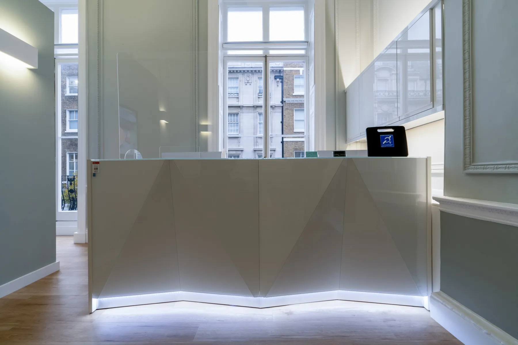
How 3Beam Imaging Centre supports your endodontic workflow
3Beam Imaging Centre, based at 86 Harley Street, London, has invested in the Morita Veraview X800 specifically for demanding endodontic and root canal indications.
For referring dentists and endodontists we offer:
-
Dedicated small-FOV endodontic CBCT protocols on the Morita X800
-
Same-day appointments for urgent root canal and retreatment cases (subject to availability)
-
Structured radiology reporting by Dr Rebecca Davies, Consultant Dentomaxillofacial Radiologist
-
Secure digital delivery of DICOM data and an easy-to-use viewer so you can review scans chairside
-
Clear referral pathways for:
-
Non-healing root canal treatments
-
Suspected vertical root fracture
-
Complex anatomy and missed canals
-
Pre-surgical endodontic planning
-
The goal is simple: to integrate high-quality Morita CBCT seamlessly into your endodontic treatment workflow, without disrupting your clinic schedule.
When to refer for a Morita endodontic CBCT scan
As a practical guide, consider referring for CBCT on a Morita X800 when:
-
You are planning complex root canal treatment on molars or teeth with suspected anatomical variation.
-
Symptoms persist after root canal treatment but 2D imaging is inconclusive.
-
You suspect a vertical root fracture or resorptive defect.
-
You are planning apical surgery near the sinus, IAN or mental foramen.
-
You are managing complex root canal retreatment cases with previous failures.
In each of these scenarios, a small-FOV Morita scan can provide information that genuinely changes your treatment plan.
Refer your endodontic and root canal cases to 3Beam
If you are a general dentist, endodontist or multi-disciplinary clinic looking for a high-quality imaging partner for your endodontic treatment and root canal cases, 3Beam can support you with:
Endodontic CBCT scans on the Morita Veraview X800, with same-day appointments where possible and formal radiology reporting by Dr Rebecca Davies.
To set up a streamlined referral pathway for your endodontic treatment and root canal patients, register with 3Beam to start using our online portal and start referring your endodontic and root canal cases directly.