
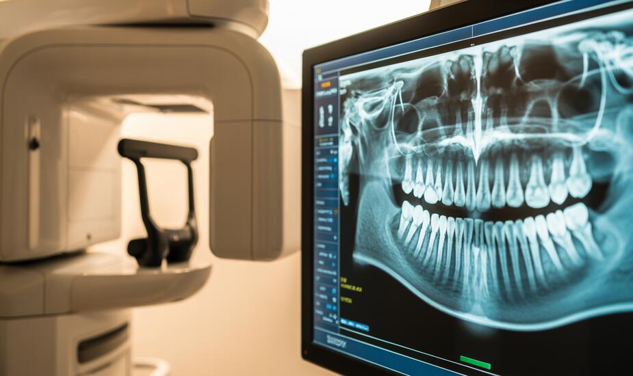
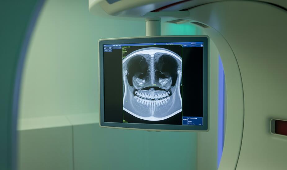
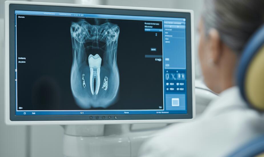
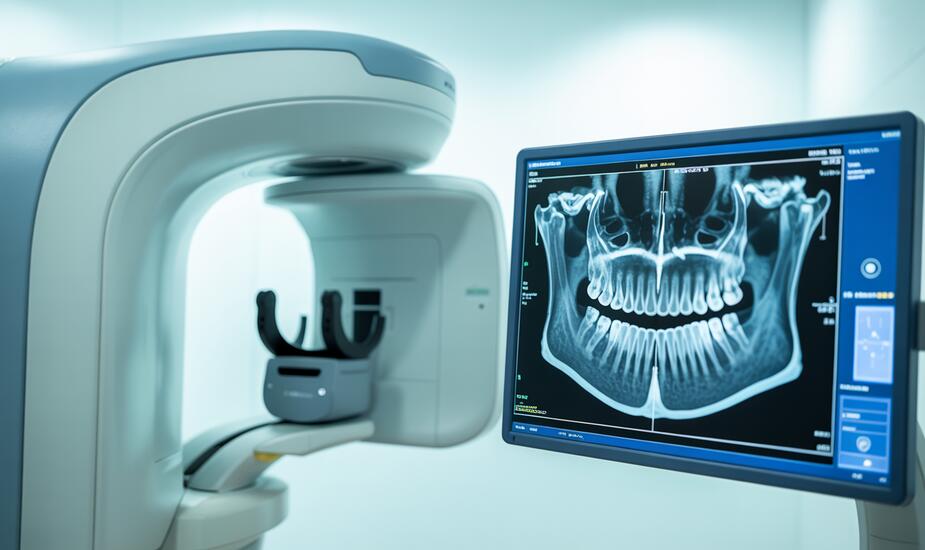
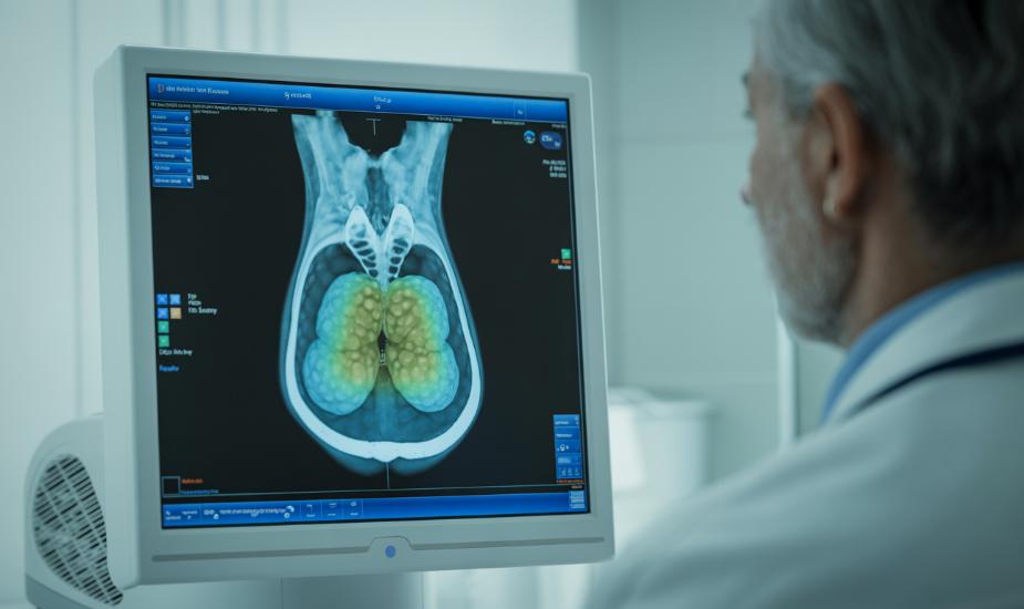
CBCT
CBCT-Guided Implant Surgery: How 3D Imaging Powers Digital Surgical Guides for Precise Implant Placement
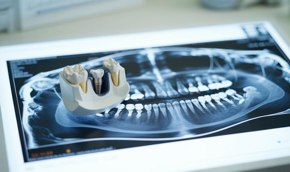
CBCT guided implant surgery delivers sub-millimetre accuracy via digital surgical guides. Same-day imaging at 86 Harley Street, London. Book now.
Read MoreCBCT for Dental Trauma: How 3D Imaging Detects Root Fractures and Guides Emergency Treatment
CBCT detects root fractures, luxation injuries and alveolar fractures missed on periapical radiographs. Same-day dental trauma imaging at 86 Harley Street, London.
Read MoreCBCT for Odontogenic Sinusitis: When Dental Pathology Causes Sinus Disease
CBCT odontogenic sinusitis imaging identifies dental causes of maxillary sinus disease. Same-day 3D scans with radiologist reporting at 86 Harley Street, London.
Read MoreCBCT for Peri-Implantitis: How 3D Imaging Detects Bone Loss Around Failing Dental Implants
CBCT peri-implantitis imaging reveals the full 3D extent of bone loss around failing implants. Book a same-day scan at 86 Harley Street, London.
Read MoreCBCT for Jaw Pathology: How 3D Imaging Detects Cysts, Tumours, and Odontogenic Lesions
CBCT jaw pathology imaging detects cysts, tumours, and odontogenic lesions with 3D precision. Same-day appointments at 86 Harley Street, London.
Read MoreCBCT for Root Resorption: How 3D Imaging Detects What Periapical Radiographs Miss
CBCT root resorption imaging detects lesions that periapical radiographs miss. 3Beam offers same-day 3D scans with radiologist reporting at 86 Harley Street, London.
Read MoreCBCT Cephalometric Analysis: How 3D Imaging Is Transforming Orthodontic Diagnosis and Treatment Planning
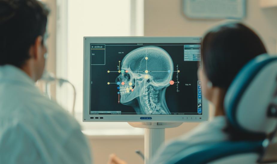
CBCT cephalometric analysis gives orthodontists 3D diagnostic precision for skeletal assessment, orthognathic planning, and growth monitoring. Book a same-day scan at 3Beam, 86 Harley Street.
Read MoreCBCT for Sinus Lift Planning: How 3D Imaging Ensures Safer Maxillary Sinus Augmentation
Need CBCT sinus lift planning? 3Beam offers same-day 3D imaging with consultant radiologist reporting at 86 Harley Street, London. Book now.
Read MoreCBCT Airway Analysis for Obstructive Sleep Apnoea: What Dentists and ENT Surgeons Need to Know
Need CBCT airway analysis for sleep apnoea? 3Beam offers same-day 3D airway imaging with radiologist reporting at 86 Harley Street, London. Book now.
Read MoreCBCT for Periodontal Bone Loss: How 3D Imaging Transforms Staging, Surgical Planning and Treatment Outcomes

CBCT periodontal bone loss imaging detects buccal and lingual defects missed by 2D radiographs. Learn how 3D imaging improves staging, furcation assessment, and surgical planning. Same-day scans at 86 Harley Street.
Read More